Ear Anatomy
The ear can be divided into three parts: the outer ear, middle ear, and inner ear.
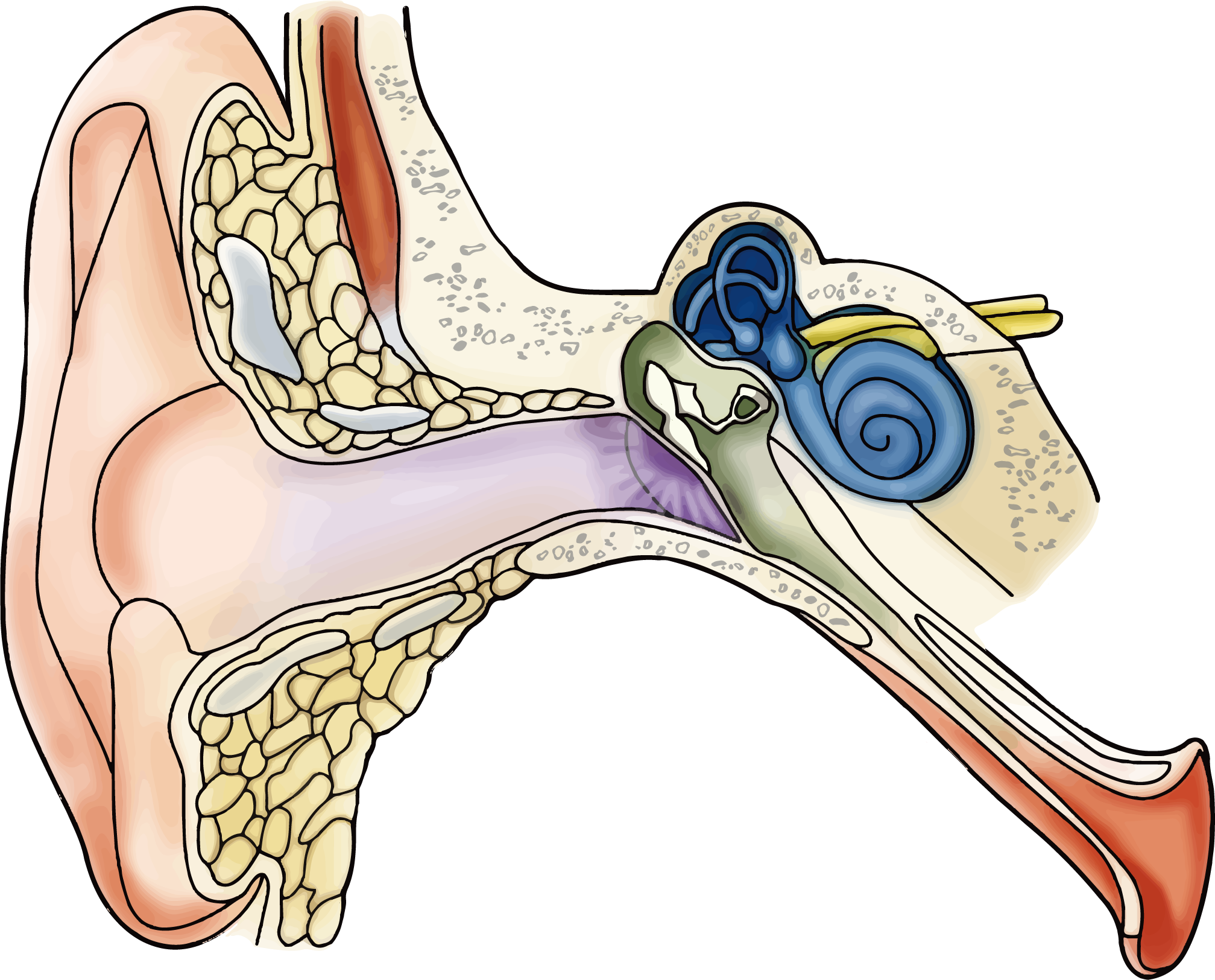
Outer ear: The internal part is higher, so examination requires lifting upward.
Middle ear: Located behind the eardrum.
Inner ear: Includes the vestibule, semicircular canals, and cochlea.
Common Ear Conditions
- Earwax impaction in the external auditory canal
- External ear infection
- Middle ear effusion
- Chronic otitis media
- Inner ear imbalance
- Sudden hearing loss
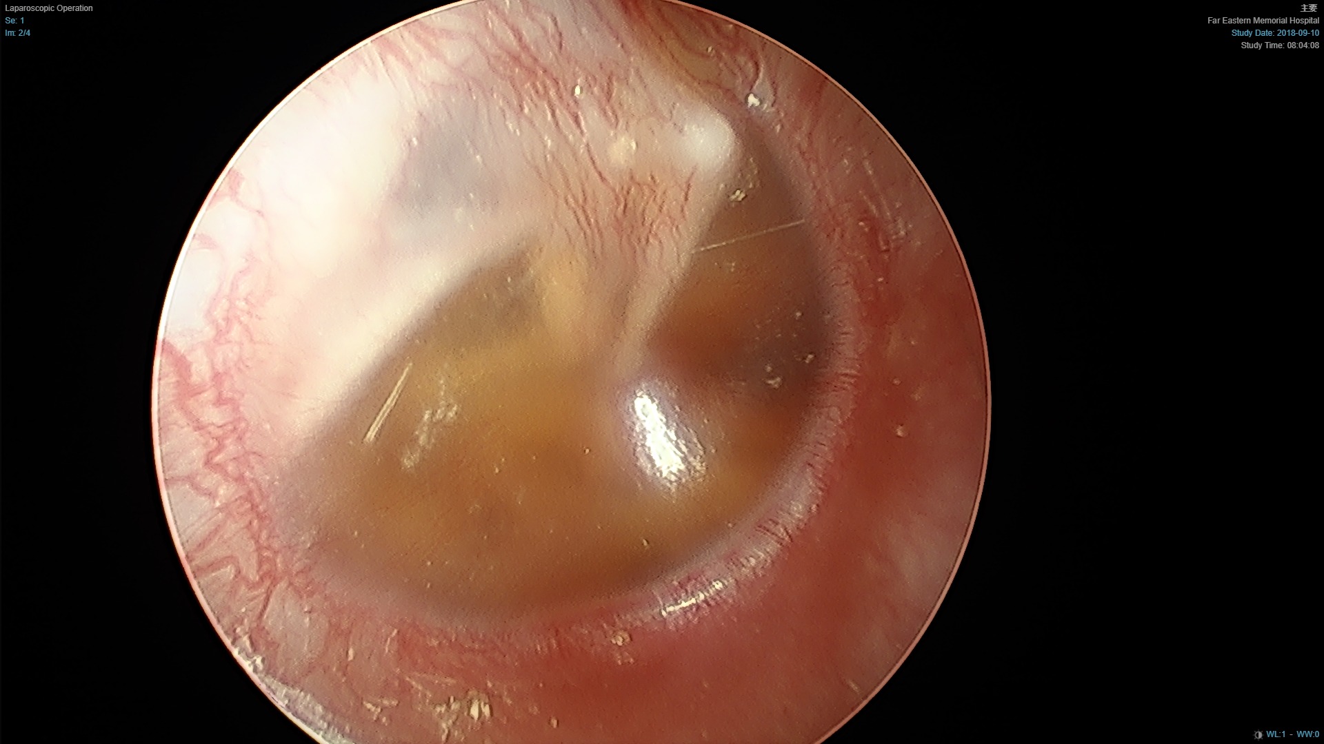
Middle Ear Effusion
If middle ear fluid persists for over three months or causes significant hearing loss, placement of a tympanostomy (ventilation) tube may be recommended.
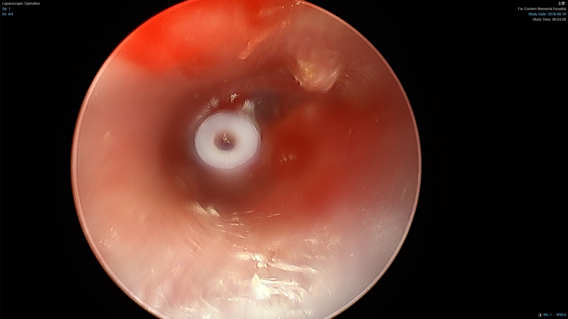
Tympanostomy Tube (approx. 2.7 mm)
The tube allows drainage of middle ear fluid, relieves negative pressure, and improves ventilation. It is button-shaped (see below). Patients do not feel it in the eardrum. The tube typically falls out naturally into the ear canal after about one year.