Neck Soft Tissue Ultrasound
Neck Ultrasound: A Valuable Tool for Evaluating Neck Masses
Neck masses can have many causes, such as thyroid nodules, lymph node enlargement, salivary gland tumors, or other rare lesions. It is recommended that an ENT/head and neck surgeon first performs a clinical assessment and physical examination to make a preliminary diagnosis. For example, nasopharyngeal cancer may be directly evaluated with a biopsy. If further evaluation is needed, the physician may arrange a neck ultrasound examination.
Features of Neck Ultrasound
- Helps the physician and patient determine the cause of the neck mass
- No radiation exposure; preferred imaging when physical exam shows no abnormalities
- Allows early diagnosis
- Facilitates appropriate treatment
Neck Ultrasound Procedure
- After history and physical exam by an ENT/head & neck surgeon, further imaging is arranged if needed
- The ultrasound room is located on the 6th floor, Endoscopic Ultrasound Center
- Due to serving over 2,000 patients per year, a 1–2 week waiting period may be required
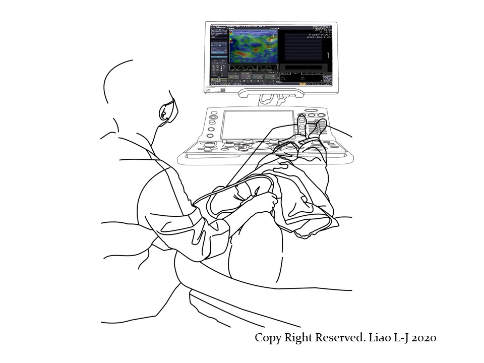
Patient Positioning During Neck Ultrasound (see right image)
- The patient lies on their back with a pillow under the shoulders
- Both sides of the neck, including thyroid, salivary glands, and lymph nodes, are routinely evaluated
- Assessment includes grayscale, color Doppler, and elastography
Typical Findings in Malignant Neck Lesions
- Irregular vascular distribution
- Disorganized ultrasound imaging
- Larger size
- Increased stiffness on elastography
- If malignancy is suspected, ultrasound-guided fine needle aspiration or core needle biopsy is recommended for pathology evaluation